LABNIRS
Toward Next-Generation Optical Brain-Function Imaging
- Описание
- Application
- About NIRS
- Performance
- Operation
- Scalability
- Configuration
Описание
Just how the human brain functions remains one of the greatest unsolved puzzles. To solve this mystery, brain-function imaging for visualization of brain functions has developed rapidly in recent years. In particular, in vivo optical imaging by functional near-infrared spectroscopy (fNIRS) has attracted attention as a technique that supports next-generation brain science. Utilizing its leading-edge science and technology, Shimadzu has developed the LABNIRS, thereby contributing to the still growing field of brain science.
High Performance
Next-generation optical brain-function measurements start withmulti-channel and high density
High-speed sampling
Reliability of 3 wavelengths and photomultiplier tube achieve superb sensitivity
Easy Operation
Intuitive user interface
Measurement and analysis by simple button clicks
Outstanding Scalability
Comprehensive options provide powerful measurement support
Increase the number of channels according to the aim of the experiments
Application
Cognitive Neuroscience
To acquire strictly task-dependent signals, the brain-function measurements are performed in a laboratory where various conditions can be controlled. fNIRS places few restrictions on the measurement environment or the subject’s posture, which makes it effective for psychological experiments. Acquiring brain-function data with respect to diverse topics to understand human cognitive functions under various conditions may lead to the kansei evaluation of products.
Eye Movements
Simultaneous measurements with an eye movement tracker can link the subject’s eye movements and brain functions.
Drug Development and Medical Research
Measurements on Newborn Babies and Infants
The brain functions of newborn babies and infants and how they develop is a research area not yet fully explained. Because it uses near infra-red light, fNIRS is a safe brain-function measurement technique effective for measurements on newborn babies and infants. It can be used for diverse research applications including the senses of touch, hearing, and vision.
Bibliography
・ NeuroImage, 54 (3), 2394-2400 (2011)
・ Brain Research, 1383, 242-251 (2011)
・ NeuroReport, 23 (6), 373-377 (2012)
Basic Research
Simultaneous NIRS and EEG Measurements
Simultaneous measurements of neural activity and changes in blood flow enhance the spatiotemporal resolution and permit application to many research fields.
Bibliography
・ Brain Topogr, 22(3), 197-214(2009)
・ NeuroImage, 59(4), 4006-4021(2012)
Informatics Research
Motion Analysis
Simultaneous measurements with a motion analysis system can link the subject’s body movements and brain functions.
Bibliography
・ NeuroImage, 34 (4), 1416-1427 (2007)
This page may contain references to products that are not available in your country.
Please contact us to check the availability of these products in your country.
About NIRS
About NIRS (Principle of Operation and How It Works)
What Is NIRS (Near-Infrared Spectroscopy)?
The human brain has about 100 billion neurons. Humans take in the information associated with sight, hearing, touch, smell, and taste from sensory organs (eyes, ears, etc.) and convert this information to electrical signals which are then sent to the brain. Neurons in the brain process this information through mutual exchange of the signals to determine the next action. During this process, oxygenated hemoglobin (oxyHb) supplies oxygen via capillary vessels. NIRS technology can analyze the functional localization of the brain by measuring this reaction in real time using near infrared light.
Principle of NIRS (Near-Infrared Spectroscopy) — Why use near infrared light?
The blood component hemoglobin scatters light, and the ratio of infrared light absorbed to that scattered changes depending on the degree of hemoglobin binding with oxygen. NIRS measures this rate of change and the change in oxygenated hemoglobin concentration.
Near infrared light having a wavelength of 700 to 900 nm is used in the optical measurement of living organisms. Visible light (wavelength 400 to 700 nm) is substantially absorbed by hemoglobin and other component organic matter, while absorption by water increases at wavelengths longer than near infrared light. Thus, light in these wavelength ranges cannot internally penetrate living organisms. Thus, the near infrared wavelength region is also referred to as «a window to living organisms» because near infrared rays easily penetrate living organisms.
Though absorption of light in this wavelength region is caused mainly by oxygenated hemoglobin (oxyHb) and deoxygenated hemoglobin (deoxyHb), both of these have differing absorption spectra, and the isosbestic point is in the vicinity of 805 nm, as shown in the figure on the right. For this reason, if the mol molecular absorption coefficient of oxyHb and deoxyHb is known, the change in oxyHb and deoxyHb concentration can be calculated by measuring the change in absorption at two or more wavelengths.
Principle of Brain Function Measurement by Near Infrared Light
Though near infrared light can penetrate relatively deeply into living organisms, it does not penetrate easily into the human head. For this reason, the inside of the brain is irradiated from the surface of the head by near infrared light via optical fiber, and light, after being absorbed and scattered at the cerebral cortex, is condensed by optical fiber again at a distance of about 30 mm from the surface of the head in the case of an adult. At this time, light reaches an area about 20 mm deep from the surface of the head, indicating that it is absorbed by hemoglobin at the cerebral cortex. Because living organisms significantly scatter light, near infrared light introduced by optical fiber is scattered by various types of tissue. Some of this scattered light reaches the optical fiber in the light receiving probe, and is then guided to a photomultiplier where it is converted to electrical signals.
This page may contain references to products that are not available in your country.
Please contact us to check the availability of these products in your country.
Performance
High Performance
Higher Temporal and Spatial Resolution
【1】 Accepts up to 40 sets––2.5 times more than before (142 channels max.).
【2】 Spatial resolution doubled for high-density measurements.
【3】 Captures rapid cerebral blood flow signals in just 6 ms (previously 25 ms).
High Sensitivity is the Basis for Reliable Measurements
【1】 Three semiconductor laser wavelengths capture more accurate data.
【2】 A photomultiplier tube captures the weakest cerebral signals.
Holder: FLASH*1 (Flexible Adjustable Surface Holder)
Flexible Adjustable Surface Holder (FLASH) supports more stable measurements.
Select from the wide range of holders, which are easily customizable.
This page may contain references to products that are not available in your country.
Please contact us to check the availability of these products in your country.
Operation
Graphical User Interface
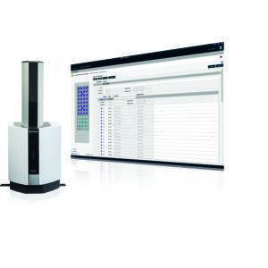
The intuitive user interface permits complex measurements and analysis conditions settings with simple button clicks.
Measurement Mode
【1】 Simple design of fiber placements
【2】 The set fiber placements are reflected on the trend graph and map during measurements.
Analysis Mode
【1】 Comprehensive data processing functions
Features a variety of analysis and processing tools, including independent component analysis (ICA*3), frequency filters, task addition, channel addition, centroid and integral analysis.
【2】 Statistical analysis functions
General linear model (GLM) statistical processing offers simple statistical analysis and evaluations at the point of measurement.
【3】Multi-distance functions
Combining short-distance measurements reduces the effects of scalp blood flow and other extra signals.
【4】Batch processing functions
Permits batch processing with predetermined analysis procedures.
【5】Data output functions
Permits output as text files (compatible with NIRS-SPM).
【6】Data continuity
FOIRE Series data can be loaded to make use of past data. *3 Patented in Japan 04379155
This page may contain references to products that are not available in your country.
Please contact us to check the availability of these products in your country.
Scalability
Options
Comprehensive options support diverse research requirements.
Video recording system
Records synchronized video images of the environment and a subject’s body movements during measurements.
Simultaneous EEG Measurement
This permits simultaneous NIRS and EEG measurements. LABNIRS high-speed sampling is effective for simultaneous EEG measurements. Please consult your Shimadzu representative.
Stimulus presentation system
This allows testing with precise control of the presentation timing of visual and voice stimuli.
3D Position Measurement System Magnetic
Attach the fibers to measure three-dimensional information. This is an indispensable item to achieve highly reproducible measurements.
Fiber Extensions
Fibers can extended up to 13 m to suit the application. Effective for measurement synchronization with MRI and for measurements inside a chamber or driving simulator.
MRI Fusion Software
Map images based on 3D information can be displayed over individual MRI images. Using a whole-head FLASH holder permits seamless mapping of the entire brain.
System Expansion
The number of the channels can be increased depending on your purpose.
The channels can be added in field by research expansion and available budget. Initial investment can be held down and upgrading by step is possible without returning the instrument to the factory.
Customization of the system with 4 sets (10 channels) to 40 sets (142 channels) is available.
Real-Time Data Transfer System
This supports biofeedback with the subject and brain-machine interface (BMI) to control external devices by transferring measured data to another PC in real time. Please consult your Shimadzu representative.
This page may contain references to products that are not available in your country.
Please contact us to check the availability of these products in your country.
Configuration
Flexible System Configuration
Three Steps to Customize the Best System for You
- Select the main unit.
Select between 4 and 40 sets in increments of 4 sets to suit your research application. More sets can be subsequently added.
- Select the fiber shape.
Select L-shaped or straight-type optical fibers to suit the research application and measurement environment.
- Select the holder.
Select the holder you need from the various types available
- Select the options.
Select the required options for your research application.
Customized Examples
Customizable for wide range of researches
Measurement of entire brain
For research of a spontaneous brain activity, various psychology and behavioral testing, etc. |
Measurement in physical exercise
For rehabilitation research, research for perception of three-dimensional space, etc. |
Simultaneous measurement of NIRS and EEG
For further research in combination with nerve activity by measuring spatiotemporal closely such as simultaneous measurement with EEG, etc. |
||||||||||||||||||
Use of real-time data
For robot control, biofeedback, etc. |
Simultaneous measurement on multiple examinees
For research on communication or decision-making, etc. Please use a Holder set for newborn babies in the case of parent and child’s communication research. |
This page may contain references to products that are not available in your country.
Please contact us to check the availability of these products in your country.


